Meiosis is the chromosomal reduction process that enables the formation of the haploid gametes (sperm and egg cells) required for sexual reproduction. This process is also crucial because it promotes genetic diversity and makes it easier to correct genetic flaws through recombination. In this article, you will learn more about the stages of cell division in meiosis and its comparison to mitosis. Also, the meiosis concept map is presented to make it easier for you to visualize the process.
Meiosis Concept Map
- Phases of Meiosis
- Involvement of Meiosis in Genetic Variation
- Key Differences between Meiosis and Mitosis
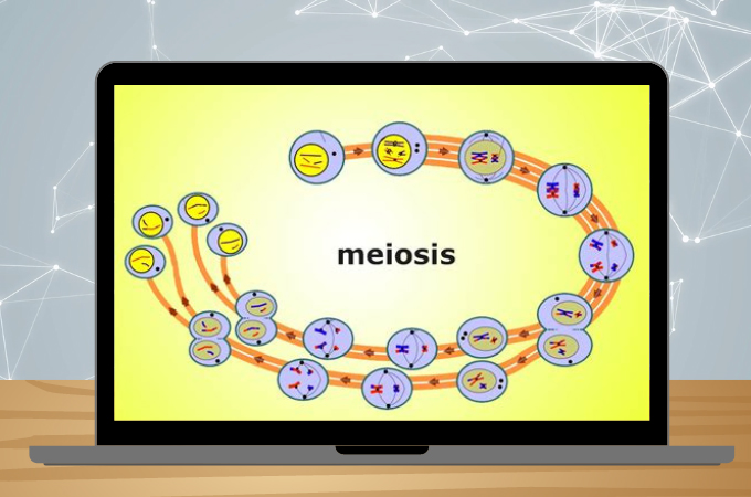
Phases of Meiosis
A two-step division procedure is used during Meiosis. During the initial round of cell division, known as meiosis I, homologue pairs split apart. During meiosis II, the second cycle, sister chromatids split. Since meiosis involves two rounds of cell division (Meiosis I and II), a starting cell can produce four gametes (eggs or sperm).
Below is the Meiosis concept map to visualize the phases and stages that occur in this cell division procedure.

Meiosis I 4 Stages of Cell Division
In eukaryotes, the prophase marks the beginning of the cell division stage. Homologous chromosomes link up and create synapses during prophase I, a process that only occurs during meiosis. The paired chromosomes are known as bivalents, and it is now clear that genetic recombination is what causes chiasmata to occur. These can be seen under a microscope thanks to chromosomal condensation.
After prophase I, individual chromosomes are typically dispersed throughout the cell nucleus. The chromosomes of the cell condense and move toward one another, aligning in the center of the dividing cell, and the nucleus of the cell disintegrates during metaphase I. Sister chromatids, or homologous chromosomes during meiosis I, split during anaphase and travel to opposing poles of the cell under the influence of microtubules. When nondisjunction occurs, the homologous or sister chromatids are drawn to one pole of the cell because the separation does not take place.
The chromosomes are encased in nuclei during telophase I, which is the last stage in meiosis I. The original cell’s cytoplasm is now split into two daughter cells as the cell goes through a process known as cytokinesis. Only one set of chromosomes, or half as many as the original cell’s total number, are present in each haploid daughter cell.
Meiosis II 4 Stages of Cell Division
The chromosomes condense and new pair of spindle fibers develop during prophase II. Moreover, the chromosomes start to move in the direction of the cell’s equator. The centromeres of the paired chromatids align along the equatorial plate in both cells during metaphase II. The centromeres of the paired chromatids align along the equatorial plate in both cells during metaphase II. The chromosomes split at the centromeres during anaphase II. The divided chromosomes are drawn toward the cell’s poles by the spindle fibers.
In anaphase II, the chromatids divide at the centromere and go to the opposite poles along the spindle fibers. Once more dividing, the cells contract in the middle. Four cells, each with half of the original genetic material, are the final outcome. For males, each cell develops into a sperm. Meanwhile, for females, one cell develops into an egg, while the other three become polar bodies that are dormant.
Involvement of Meiosis in Genetic Variation
One gamete from each parent joins together to produce a zygote during fertilization. Each gamete has a unique DNA set because of recombination and independent assortment during meiosis. Further, the resultant zygote has a special set of genes as a result. Also, the rearrangement of genes into distinctive combinations boosts genetic diversity in a population and is responsible for the differences between siblings of the same parents. At it shows in the meiosis mind map to further see how this cell division procedure increases human genetic variation.
During meiosis, a mechanism known as independent assortment causes the chromosomes to randomly travel to different poles. After meiosis, a gamete will have 23 chromosomes, but independent assortment implies that each gamete will have one of the numerous possible chromosome combinations.
Recombination or crossing over occurs during prophase I. Identical chromosomes 1 from each parent pair run the length of each gene. Along their length, chromosomes break, reassemble, and trade part of their genes. During this stage, the chromosomes possess a unique set of genes.
Key Differences between Meiosis and Mitosis

Even though mitosis and meiosis have some similarities, still, these two cell division have their key differences. Meiosis creates cells that are genetically distinct from the parent and are haploid or the halving of chromosomes. On the other hand, in the mitosis procedure, the daughter cells are diploid and identical to the parent cell. With this idea, it is apprehended that meiosis and mitosis are both undergoing DNA replication. And what makes them different from each other is that mitosis generates new body cells, while meiosis develops sperm and egg cells. The mitosis concept map is also prepared below in order for you to see how it works and to easily compare it with meiosis.
Conclusion
It seems that studying meiosis may include phases that are complicated, but with the meiosis concept map, it is now more convenient to comprehend each stage. In addition to this, the comparison of meiosis and mitosis is easier to visualize using a mind map. Learning the process is easy to understand with concept maps, so if you want more like these stay tuned for the next blog.
Leave a Comment